

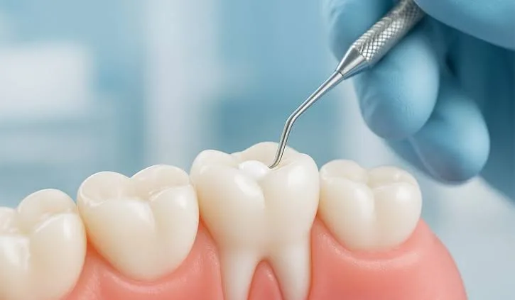
The oral cavity, in addition to being the starting point of the digestive system, is also at the center of speech function and facial aesthetics. A healthy enamel layer is the hardest tissue in the body and the most resistant to external factors. However, over time, dietary habits, inadequate oral hygiene, and acidic changes in the oral flora can compromise the integrity of this strong structure. One of the most basic protective and restorative procedures in dentistry, filling treatment refers to the reconstruction of lost tooth tissue with biocompatible materials.
If you are experiencing structural loss in your teeth, sensitivity to hot and cold, or pain while chewing, and you are researching filling treatment in Bursa at the clinical level, this comprehensive guide will allow you to objectively examine the medical background of restorative procedures and current treatment protocols.
This article has been prepared in accordance with Law No. 1219 and health services information regulations, with the aim of informing the public about oral and dental health practices.
How Does the Tooth Decay (Caries) Mechanism Work?
Tooth decay is not a condition that appears suddenly, but a gradual demineralization (mineral loss) process. Bacteria such as "Streptococcus mutans," naturally found in the oral flora, metabolize the carbohydrates and sugars we consume and release lactic acid as a by-product. These acids begin to dissolve the calcium and phosphate crystals on the outermost surface of the tooth.
Initial (Enamel) Lesion: It begins as chalky white spots in the enamel layer. At this stage, the tooth has not yet formed a cavity (hole), and the process can be stopped with good oral care.
Dentin Caries: Bacteria that pass through the enamel layer reach the dentin layer, which has a softer and more porous structure. Since dentin contains microscopic tubules connected to nerve endings, patients begin to feel sensitivity to sweet, hot, or cold stimuli at this stage.
Advanced Destruction: If decay that spreads rapidly in the dentin layer is not cleaned, it reaches the pulp (nerve) tissue, considered the heart of the tooth, causing infection and severe nighttime pain. This condition exceeds the limits of filling treatment and requires root canal treatment.
In Which Indications Is Filling Treatment Used?
Although it is commonly thought that fillings are applied only for bacteria-related decay, restorative procedures come into play in many different clinical situations that cause tissue loss in dentistry:
Traumatic Fractures: Breaks occurring in the crown part of the tooth as a result of impact, a fall, or biting a hard object.
Abrasion: Wedge-shaped tissue losses created at the gum line by aggressive brushing with hard-bristled brushes or abrasive (gritty) toothpastes.
Attrition and Bruxism: Flattening and enamel cracks that occur on the chewing (occlusal) surfaces of teeth due to the habit of grinding teeth at night (bruxism).
Erosion: Chemical dissolution on the enamel surface as a result of frequent consumption of acidic drinks or stomach acid reaching the mouth (reflux/bulimia).
Secondary Caries: New caries formations progressing beneath old fillings placed years ago, where the marginal fit has deteriorated and leakage has begun.
Materials Used in Restorative Dentistry
In the clinical processes of filling treatment in Bursa applied in modern dentistry, the choice of material is determined by the dentist according to the extent of tooth substance loss, the aesthetic needs of the area, and chewing forces. The main materials used in line with biomimetic (nature-mimicking) approaches are as follows:
Composite Resins
These are polymer-based restorative materials reinforced with glass particles that can precisely mimic tooth color and translucency. They not only adhere to the tooth mechanically, but also integrate with tooth tissue thanks to chemical bonding agents (adhesion). They are safely used in anterior aesthetic restorations and posterior chewing teeth. With special light devices (polymerization), they harden within seconds and gain their final strength.
Indirect Restorations (Inlay, Onlay, and Overlay)
These are laboratory-supported porcelain restorations preferred when the damage in the tooth is too extensive to be repaired with a standard composite filling, but too small to require full coverage of the tooth (crown). After the decay is removed, a digital impression of the tooth is taken. Produced in a laboratory from ceramic or reinforced composite blocks, these pieces are bonded to the tooth with high precision like a missing puzzle piece. They are exceptionally durable against chewing forces and carry a very low risk of microleakage.
Glass Ionomer Cements
These are highly biocompatible materials capable of releasing fluoride. Since they are not as resistant to chewing pressure as composites, they are generally used in pediatric dentistry (primary teeth), root surface caries, or as a protective base material beneath permanent composite fillings.
Clinical Procedures: Step-by-Step Filling Treatment
The long-term service life of a healthy filling depends on the precise clinical protocols to be applied by the dentist.
1. Examination and Anesthesia: The procedure begins with a radiographic examination of the relevant tooth. After the boundaries of the decay are identified, local anesthesia is applied so that the patient feels no pain.
2. Isolation Process: For composite resins to bond strongly to the tooth, the working area must be completely dry (free of saliva, blood, or breath moisture). At this stage, a rubber sheet (rubber dam) is placed around the tooth to provide complete isolation.
3. Excavation (Cavity Preparation): Using rotary instruments and hand tools called excavators, the infected, softened decayed tissue is completely removed until the healthy enamel and dentin boundaries are reached.
4. Surface Roughening and Bonding (Adhesion): An acid gel (phosphoric acid) is applied to create microscopic pores on the tooth surface. Then the bonding resin (bond), which seeps into these pores and forms a bridge between the tooth and the filling material, is applied and light-cured.
5. Layering Technique (Incremental Placement): The composite filling material is placed in thin layers rather than as a single mass in the prepared space. Each layer is light-cured separately. This method minimizes the material's shrinkage risk, preventing the stress (post-operative sensitivity) that the filling could create in the tooth.
6. Polishing and Occlusion Check: After the cusps and grooves are shaped in accordance with the tooth's original anatomy, the height is checked. Early contact points are reduced to ensure harmonious closure of the upper and lower teeth. In the final stage, the filling surface is polished with special rubber and paste until it reaches glass-like smoothness.
Post-Procedure Adaptation and Oral Care
After the clinical procedures are completed, maintaining oral health is the patient's responsibility. The following medical rules are recommended during the healing and adaptation period:
Food and drink should be avoided until the effect of local anesthesia (numbness) has completely worn off (approximately 2-3 hours). The feeling of numbness can cause deep tissue injuries due to involuntary biting of the cheek or lip.
Mild cold-hot sensitivity in the tooth during the first days after treatment is a physiological adaptation process and usually decreases and disappears within a few days. If the sensitivity becomes more severe or lasts for weeks, a dentist's evaluation is required.
The belief that a tooth that has undergone filling "will no longer decay" is medically incorrect. Microscopic boundaries where the filling margins meet the natural tooth carry a risk for secondary caries if brushing is inadequate. Therefore, brushing at least twice a day with fluoride toothpaste and regular floss use directly determine the clinical lifespan of the filling.
Repairing early-diagnosed caries with restorations performed in accordance with biological principles is the most fundamental step in preventing tooth loss. For accurate diagnosis and personalized treatment planning, having an examination at a well-equipped dental clinic is the most reliable approach.
Legal Notice and Information
This page content is intended solely to inform the public about preventive and restorative dentistry within the framework of the Law No. 1219 on the Practice of Medicine and its Branches and the relevant Regulation on Promotional and Informational Activities in Health Services. The information provided herein cannot be used for the diagnosis or treatment of diseases, does not offer any guarantee of success, and is not intended for commercial promotion. To ensure the most appropriate treatment plan for your oral and dental structure, you must consult a dentist directly for an examination.