

Teeth Whitening (Bleaching) Procedures: Chemical Mechanism, Clinical Protocols, and Indications
The natural color of the tooth emerges as a result of the optical interaction between the light-transmitting semi-transparent enamel layer and the dentin tissue beneath it, which gives the tooth its main yellow-brown tone. Over time, organic and inorganic pigment accumulation occurs in these tissues due to various intrinsic and extrinsic factors. In clinical practice, the protective treatment protocol we apply to lighten tooth color solely through the principle of chemical oxidation, without any mechanical abrasion of the tooth tissue, is called teeth whitening (bleaching). This procedure is a medical method used in cases where the natural tooth color achieved after tartar cleaning (detartrage) and polishing is insufficient.
Etiology of Tooth Discoloration and Clinical Classification
The success of whitening treatment depends directly on correctly diagnosing the source of discoloration. In clinical examination, we evaluate discolorations in two main groups:
Extrinsic Discolorations: These are pigments that adhere to the pellicle layer on the enamel surface as a result of the use of chromogenic substances such as tea, coffee, tobacco products, or long-term medicinal mouthwashes containing chlorhexidine. While superficial stains are removed by polishing, whitening agents are needed for persistent pigmentation that penetrates the enamel prisms.
Intrinsic Discolorations: These are deep color changes incorporated into the enamel and dentin structure during tooth development or after eruption. This group includes tetracycline antibiotics used during childhood, dental fluorosis caused by high fluoride levels in drinking water, the thinning of the enamel layer with age and the increased prominence of the underlying yellow dentin, and tissue hemorrhage products in teeth that have lost vitality (necrotic) as a result of trauma.
Biochemical Mechanism: How Do Whitening Agents Work?
In tooth whitening, hydrogen peroxide or carbamide peroxide gels that break down in the mouth and convert into hydrogen peroxide are used as the main active ingredients. Enamel and dentin tissue have a semi-permeable structure at the microscopic level.
Free oxygen radicals released from the peroxide-based gel applied to the tooth surface pass through the spaces between enamel prisms and reach the dentinal tubules. There, they oxidize the high-molecular-weight, dark-colored organic ring compounds (chromophores) that cause discoloration. As double carbon bonds break and turn into single bonds, these pigments break down into colorless, small molecules that do not absorb light. During the procedure, there is no material loss or abrasion in the inorganic crystal structure of the tooth (hydroxyapatite); only the optical reflection characteristics of the pigments within the tissue are altered.
Clinical Application Methods and Protocols
Three different whitening protocols are applied depending on the patient's tooth structure, the type of discoloration, and the clinical indication:
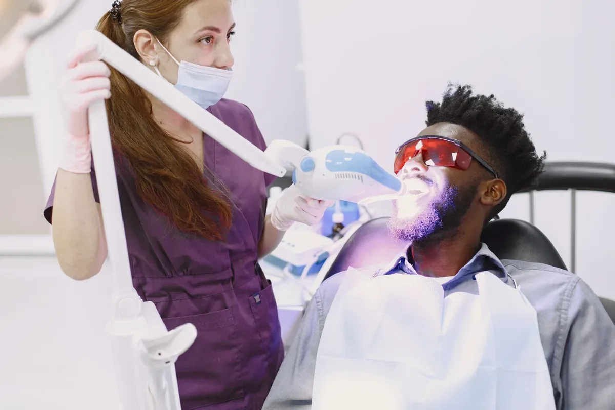
Office Bleaching: This is a procedure performed directly in the clinic by the dentist. Before the procedure, a light-cured barrier (gingival protector) is applied to the gum margin to protect the soft tissues from chemical irritation. Then, hydrogen peroxide gel in the 25% to 40% concentration range is applied to the tooth surfaces. Depending on the agent, activation is achieved chemically or with LED/laser light sources. The process is completed by performing 2 or 3 applications in the same session at approximately 15-20 minute intervals.
Home Bleaching: Custom transparent thermoplastic trays are produced using impressions taken from the patient. Lower-concentration gels containing 10% to 16% carbamide peroxide are placed into these trays by the patient and applied for 4-8 hours a day (mostly during nighttime sleep). The average treatment duration is 10-14 days. In resistant cases, office and home applications are planned sequentially (combined whitening).
Internal Bleaching (Devital Bleaching): This is applied to single teeth that have undergone root canal treatment but have turned gray over time due to tissue remnants or bleeding remaining in the pulp chamber. An access cavity is created from the back surface of the tooth, the root canal filling is isolated with a glass ionomer base, and sodium perborate or devital whitening gel is placed in the pulp chamber and sealed with a temporary filling. The agent is renewed in 3-5 day sessions until the desired shade is reached.
Contraindications, Sensitivity Management, and Maintenance Process
The whitening procedure is not applied until these pathologies are resolved in individuals with deep cracks on the enamel surface, exposed root surfaces (cementum tissue) due to gum recession, active caries, or untreated gum disease. In addition, the procedure is postponed in women during pregnancy and breastfeeding, as well as in young individuals whose enamel development has not been completed. Whitening agents act only on natural tooth tissue; they do not lighten the color of existing composite fillings, porcelain veneers, or zirconia crowns.
During or within the first 24-48 hours after the procedure, temporary sensitivity to cold and hot stimuli may develop due to fluid movement in the dentinal tubules, which is a physiological condition. To control this condition, desensitizing agents containing potassium nitrate, fluoride, or nano-hydroxyapatite are applied to the tooth surfaces immediately after the clinical procedure.
After whitening, due to the temporary water loss (dehydration) of the enamel tissue, the tooth remains highly permeable to external factors. During the first 48-72 hours, when the tissue regains water (rehydration), following the "white diet" protocol by avoiding staining factors such as tea, coffee, tomato paste, sour cherry juice, and tobacco products is essential to preserve the achieved shade.