

Root Canal Treatment (Endodontics) Procedures: Clinical Protocols, Diagnostic Criteria, and Biological Healing
When the anatomical structure of the tooth is examined, protective enamel is found on the outer surface and the hard tissue of dentin beneath it, while in the innermost layer there is connective tissue called the "pulp." This tissue, composed of blood vessels, nerves, and defense cells, provides dentin production during the tooth’s development process, and in the mature tooth it is responsible for nourishment and sensing external stimuli (pain, pressure, heat). When the pulp tissue becomes inflamed or loses its vitality as a result of various pathological factors, the clinical procedure we apply to save the tooth from extraction and keep it functionally in the dental arch is called root canal treatment (endodontic treatment).
Pulp Pathologies and Clinical Diagnostic Criteria
Healthy pulp tissue is isolated from oral bacteria as long as the enamel and dentin barriers remain intact. However, bacteria reach the pulp chamber as a result of deep dental caries, secondary leakage developing at the margins of old fillings, cracks in the teeth, or acute trauma (blow, fall).
The main conditions for which we establish an indication for root canal treatment during clinical examination are as follows:
Irreversible Pulpitis: A spontaneous throbbing pain that begins with cold or hot stimuli and continues for minutes even after the trigger is removed, especially pain that wakes the patient from sleep at night. At this stage, the inflammation in the pulp tissue has lost its ability to heal on its own.
Pulp Necrosis and Apical Periodontitis: This is the progression of inflammation to the complete loss of vitality of the nerve-vessel bundle inside the root (necrosis) and the spread of infection from the root tip (apex) to the jawbone. At this stage, hot-cold sensitivity in the tooth disappears; however, severe pain during chewing or when the tooth is touched (percussion), a lesion due to bone destruction at the root tip, and sometimes a pimple-like drainage path of infection on the gum (fistula opening) are observed.
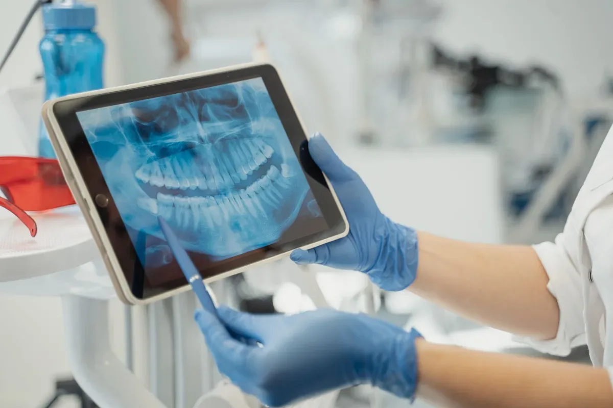
A definitive diagnosis is made by jointly evaluating intraoral vitality tests, percussion-palpation examination, and periapical radiographs showing the tissues around the root tip.
Endodontic Treatment Stages and Biomechanical Protocol
Root canal treatment is not merely the removal of nerves from the root canal system; it is the process of shaping and disinfecting it at a microscopic level. The treatment process is carried out under local anesthesia in four basic stages:
Isolation and Access Cavity: After the relevant tooth is numbed with anesthesia, the tooth is isolated with a "rubber dam" (rubber sheet) to prevent bacteria in saliva from entering the root canals. The carious tissue is completely removed to gain access to the pulp chamber.
Working Length Determination and Mechanical Shaping: The length of the root canals is measured millimetrically with the help of electronic apex locators and radiographs. Then, using flexible nickel-titanium (NiTi) rotary file systems, the infected canal walls are scraped and the canals are enlarged into a tapered form all the way to the root tip.
Chemical Irrigation: Because root canals have complex lateral branches and dentinal tubules, mechanical filing alone is not enough to eliminate bacteria. Throughout the procedure, the canals are irrigated with sodium hypochlorite (NaOCl), which dissolves organic tissue and has antibacterial properties, and EDTA solutions that remove inorganic deposits. In cases with active and resistant infection at the root tip, treatment may be divided into two sessions by placing calcium hydroxide-based antiseptic medication inside the canal.
Obturation: The cleaned, shaped, and dried root canals with paper points are filled all the way to the root tip without leaving any space (hermetic) using "gutta-percha" and tissue-friendly canal sealers (resin- or bioceramic-based sealers), which are biocompatible materials.
Coronal Restoration and Biomechanical Protection
The long-term success of a tooth that has completed root canal treatment depends not only on the root canal filling but also on the seal of the coronal restoration placed over it. During the treatment process, the loss of sound tooth walls due to caries makes the tooth fragile against chewing forces.
While direct composite resin fillings are sufficient for teeth with limited material loss, in posterior teeth that have lost most of their walls, "fiber post" applications supported by the root canal, porcelain onlay/endocrown restorations, or full zirconia crowns should be preferred in order to prevent vertical root fractures.
Healing Process, Follow-Up, and Retreatment
During the first 3 to 7 days following treatment, it is physiologically normal to feel mild to moderate pain, especially when pressure is applied, due to the healing reaction in the tissues around the root tip. This process is controlled with anti-inflammatory painkillers prescribed by the physician; if the patient does not show systemic signs of infection such as fever or widespread swelling in the facial area, there is no need to use antibiotics.
In teeth with a large bone lesion at the root tip, the regeneration of bone tissue is monitored with control X-rays taken at the 6th and 12th months. In teeth that were treated in the past but have become reinfected due to loss of the upper filling, new caries formation, or anatomical variations, direct extraction is not performed; first, the "retreatment" protocol, in which the old canal filling is removed and the canals are disinfected again, or the "apical resection" procedure, in which the root tip is surgically intervened, is evaluated.